Solo dentist
You spend 20min explaining a treatment and the patient decides later.
Show the simulation on the spot. Close more cases first visit. No need to invest in intraoral camera.

Smile simulation in seconds to close more cases in the first visit. AI-assisted radiograph analysis with hot spots. All inside the chart, with full legal audit trail.
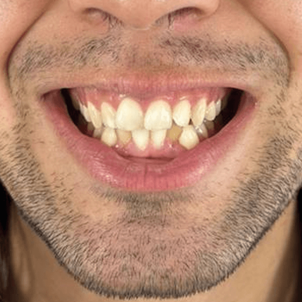
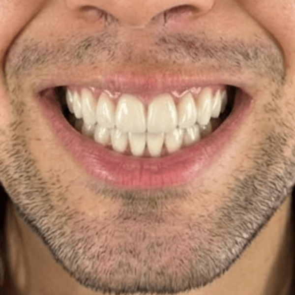
Caries · Lesions · Ceph Ricketts/Steiner
Front photo of the patient for smile, or radiograph (PNG/JPEG/WEBP) for analysis.
Gemini reads the image in seconds. Multi-treatment (veneers + whitening + orthodontics) in a single request.
Result inside the chart, with AI confidence score and automatic medical-legal disclaimer.
Side-by-side display, saved in the case and auditable. Optional digital acceptance on the spot.
Drastically increases first-visit case acceptance. Gemini Flash 3.1 model with dynamic prompt that combines multiple treatments (veneers, whitening, orthodontics) into a coherent simulation.
Assisted detection on panoramic, cephalometric, bitewing and periapical radiographs. AI flags visible findings with transparency on confidence · clinical decision is always yours, with full legal audit trail.
You spend 20min explaining a treatment and the patient decides later.
Show the simulation on the spot. Close more cases first visit. No need to invest in intraoral camera.
Every dentist reads radiographs their own way.
AI standardizes initial diagnosis. Legal audit of who saw what. Cases shared between providers.
No clinical standard across sites · each location with its own quality.
Same AI model across all locations. Single standard for simulation and analysis. Comparable acceptance metrics.
| Without visual AI | With SuperClini AI | |
|---|---|---|
| 1st-visit acceptance rate | Patient will think about it and rarely returns. | Patient sees the result and decides on the spot. |
| Explanation time per case | 15-25min with generic photos and pen drawings on the chart. | 3min showing personalized simulation. |
| Radiograph analysis | Isolated clinical eye, no second opinion. | AI flags visible findings, you decide with more context. |
| Legal audit | Manual note in chart (when remembered). | Automatic trail: who saw, when, with which AI model. |
No. AI assists diagnosis by flagging visible findings in the image, caries at stage X, suggested lesion at site Y. Clinical decision is always the dentist's. Every analysis is logged with full legal audit: who saw, when, with which AI model.
Front photo of the patient (smiling, natural open mouth). PNG, JPEG or WEBP. AI generates a 1024×1024 image showing the simulated result with selected treatments (veneers, whitening, orthodontics · combined or isolated).
Typically 30-60 seconds. Max 120s timeout with 2 automatic retries. If the model is under load, expect a small queue. You're not stuck on the screen, the result appears in the patient case when ready.
Panoramic, cephalometric, bitewing and periapical. Formats: PNG, JPEG, WEBP. Native DICOM not yet supported, for CBCT 3D, use the integrated DICOM 3D viewer (which is manual, not AI-driven).
It is not monthly, it is annual: the membership includes 20 AI patients per year, with all of AlicIA's functions on every patient you activate. When you need more, you add a single patient or a pack of 20.
No. Every simulation includes an automatic medical-legal disclaimer: 'Educational simulation · real result depends on biological and technical variables'. Use it to align expectations, never as a contract or clinical promise.
Yes. Images are sent for processing via encrypted API (TLS), we don't train public models on your data, and they're stored isolated per organization. Full legal audit per analysis.
No. AI needs connection to process. But the flow is async: you upload the image, close the screen if you want, and the result appears in the case when ready.
Full AI included in the annual membership. Guided setup in 1 business day.